The doctor used slippers to save a patient's life
The decay in Nigeria's health sector has been brought to the fore after a doctor narrated how he saved a patient's life using bathroom slippers. A Nigerian doctor, Chibuike Joseph Chukwudum has given hints on how he used a bathroom slippers to save the life of an accident victim.
Chibuike who had to improvise due to non-availiability of the materials at the hospital where he works narrated in details all he had to do to save the victim's life.
The unavailability of the needed materials many believe is a pointer to the decayed level of our health sector.
The Federal Government has been blamed for this even as Nigeria's number one citizen, President Muhammadu Buhari continues to patronize foreign medical hospitals because of the inefficient ones we have in the country.
Read Chibuike's full post below:
Just In Case You Find Yourself In A Jungle.
Emergency medicine is as exciting as it is unforgiving. People who excell in it are those who can think fast on their feet, and those who are daring. This is so as every minute counts, and the slightest delay could spell doom for the patient.
More often than not, especially in a third world country like Nigeria, the need arises for an emergency physician to reach into the deepest recesses of his brain and pull out an improvisation-- a hack, to save a patient whose life is hanging by a thread.
For people working in standard hospital settings-- where emergency tools are just a tray or trolley away-- there may not be much need to familiarize oneself with the different hacks in emergency medicine. However, for the NYSC medical officer who may find himself in a rural setting, far removed from civilization, being familiar with these hacks is very important for two reasons:
- when in a charted territory, using a hack one is already familiar with saves both oneself's time, and most importantly the patient's time.
- it has a way of sparking one's own genius to come up with a denovo improvisation when in an uncharted territory.
This is why I always share my own hacks, as I believe that it may someday be of help to someone who may find himself in a situation similar to the ones I often find myself in. As a matter of fact, I've had several colleagues call to tell me that they were in the doldrums one time or the other, and had to employ one of the several improvisations I write about, and it worked.
📝 BLUNT CHEST TRAUMA and C-SPINE INJURY, following RTA [Road Traffic Accident]
I was already on my way to work when they informed me that men of the Nigerian Road Safety Commission had just brought two accident victims to our hospital. So, I quickened my steps, knowing full well that a minute could make a world of difference.
I have a knack for smelling danger. So, immediately I entered into the mini theater, where they were being attended to, I was alarmed as I noticed that one of them was practically choking-- he was struggling to breathe and was not doing a good job about it.
My argus eye caught two things in an instant: he was coughing up blood, and the left side of his chest looked bigger than the right, and wasn't moving with each intake of breath as it should. Given the hemoptysis [coughing up blood], that could only mean that he'd taken some pretty bad hit to the chest, contused [bruised] the lung, and air was leaking freely into the pleural space, suffocating him. The fact that he sounded confused, even with no evidence of head injury, only told me how serious it must have been, as confusion in this setting is an indication of cerbral hypoxia--that the brain isn't getting enough oxygen.
I gloved up, and just like I had expected, he'd fractured multiple ribs, and there was widespread crepitus over the left side of chest, showing that there was an air leak. The trachea and the heart were pushed to one side; and air wasn't entering into his left lung at all, making the oxygen saturation to be as low as 60%. Tension pneumothorax!
Also, the cervical spine was a tad unstable, and if not attended to urgently, he could become paralysed in all for limbs. What more, the motor control of the diaphragm could also be knocked off, leading to paralysis of that muscles, and inability to breathe "at all at all," and of course death.
Bottom line? He needed two things urgently:
1. Stabilization of the cervical spine [neck bone].
2. Relieving the pneumothorax.
To be able to do number 1, above, I'd need a cervic collar. And for number 2, I'd need a wide bore needle, and then a chest tube thereafter.
However, none of these were handy, nor could they be gotten within a 100km radius.
📝 CERVICAL COLLAR HACK
Back in school, they had hammered the differemt ways of improvising a cervical collar into our heads, considering how important the cervical spine is to survival. However, I've never had cause to use any of them.
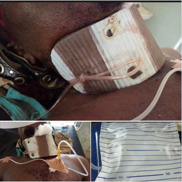
One of the hacks involves using a bathroom slipper, I remembered. So, looking around, I saw one lying carelessly. I reached for it, removed the straps, and then created a hole at the heel end, to mirror the one at the toe end. Next, I wrapped the slipper around his neck-- with soft pads underneath as a cushion; and held it in place with a tourniquet passed through the holes at both ends.
And there it was: a cervical collar!
I thanked my trauma teachers silently under my breath.
📝 NEEDLE THORACOSTOMY HACK
When someone has a tension pneumothorax, the pressure building up in his chest needs to be relieved fast, so he can breathe fine, else he could die within minutes.
Usually, tension pneumothorax is relieved by emergency needle thoracostomy, which is practically inserting a special, wide bore needle into the person's chest so that the air trapped there in, under high pressure, can escape. Visualize this as puncturing and deflating a football, or a tyre.
After this-- relieving of the tension pneumothorax by emergency needle thoracostomy-- depending on the cause, a chest tube should be passed to continue the drainage of the excessive air accumulating in the chest, else it could build up again, leading to another tension pneumothorax.
In the index case, none of the above was available-- not a standard needle for thoracostomy, nor a chest tube.
What I did was to relieve the tension pneumothorax with the regular needle that comes with syringes. Once I stuck it into his chest, you could hear jets of air escaping under high pressure. Within seconds to minutes, he began to breathe easy, and his oxygen saturation appreciated exponentially. That was the easy part.
The difficult part was how to pass a chest tube to prevent an inevitable reaccumulation of excessive air, and a recurrence of tension pneumothorax. Like I mentioned earlier, there was no chest tube available. However, a senior colleague had taught me how to improvise one by fenestrating an NG tube, or a urethral catheter, and inserting either of them into the chest. Unfortunately, none of these were available too. And moving the patient-- without knowing the full extent of his cervical injury, and not having a ready means of transportation that could carry him in one piece, without unnecessary bending of the spine-- was not an option, as it could have him paralysed in all four limbs, or worse still-- dead from paralysis of the diaphragm.
While I was still thinking of what to do, he started deteriorating again: apparently enough air had accumulated in his chest again to impair breathing, leading to hypoxemia, and malfunctioning of the brain from lack of oxygen.
Then a bright idea popped into my head. I got a wide bore cannula, and stuck it into his chest, so air could be leaving through it. Then I connected one end of a drip-giving set to the cannula, and the other end of it to a urine bag. It was an airtight arrangement.
Almost immediately, you could see mist forming along the length of the tubbing of the drip-giving set, showing that humidified air was making its way through it. After few minutes, the hitherto collapsed urine bag started inflating, showing that air was collecting there.
The challenge was how to ensure unidirectional flow of air, since there was no underwater seal to create a a valve mechanism. Without a system of valve, air could potentially flow backwards into the chest, especially when the pressure in the bag becomes higer than that in the chest.
I did two things to prevent this from happening:
1. Knowing full well that expired air is denser than atmospheric air, I made sure that the urine bag is placed at a level below the chest.
2. Most importantly, I made sure that air didn't accumulate in the bag under pressure. Once a sizeable volume of air accumulates there, we would occlude the drip-giving set with it its regulator, maintaining an air-tight system upstream. Then we would open the valve at the tail end of the urine bag, to allow the accumulated air to escape, without the risk of having atmospheric air rushing into the chest.
We carried on like this until he was fit enough, and the family was buoyant enough, to take him to a proper Hospital. 24 hours it took. And guess what. Within that period of time, there was no single incident of deterioration from hypoxemia. He was lucid all through, and breathing relatively fine.
😀
Now you have a new air-bending skill to add to the ones you already know.
I am CJ, the last air bender!
😜
📝 PS:
Needless to say, this should only be tried when there's no chest tube available; no NG tube, or urethral catheter, to improvise with; no nearby Hospital to refer the patient to [or moving the patient has its own risk]; and the BENEFITS CLEARLY OUTWEIGHS THE RISKS!
📝 NB:
Patient waa fully conscious when the pictures were taken. The purpose was explained to him, and he gave his consent.
Read post below:
No comments:
Post a Comment